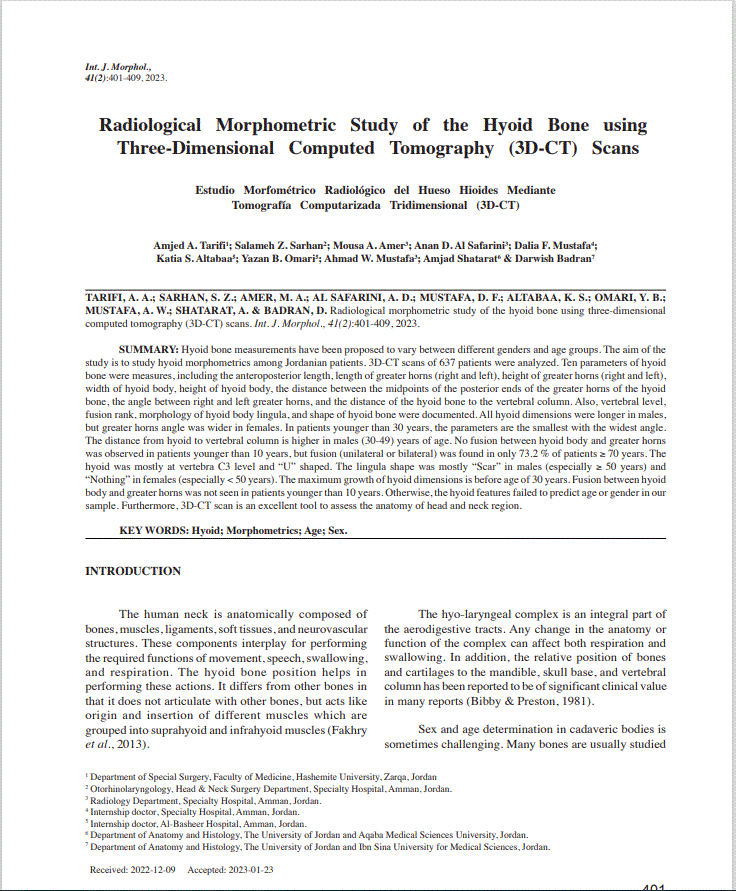
A scientific study was conducted at the Specialty Hospital on the morphology of the hyoid bone using 3D-CT imaging for both sexes and in different age groups. The study was published in one of the most important medical journals and websites on morphology "The International Journal of Morphology".
The study team consisted of a number of physicians in different specialties, Dr. Amjad Al-Tarifi, ENT Consultant, Dr. Anan Al Safarini, Radiology Resident, Dr. Musa Amer, Radiology Resident, Dr. Ahmed Mustafa, Radiology Resident, and Dr. Salama Sarhan, ENT Resident.
The study aimed to clarify whether the hyoid bone differs according to sex or age group, and whether it can be used in determining gender and age group. Noting that the hyoid bone has a vital role in breathing, speaking, and swallowing.
A sample consisted of 637 cases from the Jordanian population, and the analysis parameters focused on the dimensions and parameters of the hyoid bone in four main age groups.
In conclusion: hyoid bone's dimensions and parameters are larger in males except for the angle between greater horns which is wider in females. The maximum growth of these dimensions is before age of 30 years. In addition, fusion between hyoid body and greater horns was not seen in patients younger than 10 years. Otherwise, the hyoid features failed to predict age or sex in the sample studied. Furthermore, 3D-CT scan is an excellent tool that can be used in future studies to assess the functional anatomy of head and neck region.
Noting that the Specialty Hospital is a teaching hospital, and the hospital's healthcare staff participate in many medical researches and scientific studies that are published in international journals.
To view the study, click here